

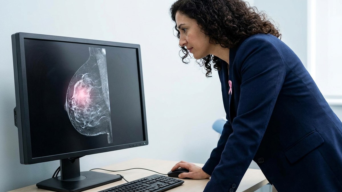
Predicting whether breast cancer has reached the axillary lymph nodes before surgery is one of the most delicate decisions in treatment. A meta-analysis published in European Radiology reviews whether artificial intelligence models applied to magnetic resonance imaging and ultrasound can do so reliably enough.
What was known
Axillary lymph node involvement is a key prognostic factor in breast cancer. Sentinel lymph node biopsy remains the reference standard, but conventional imaging depends on the radiologist’s expertise and its diagnostic accuracy varies. Recent years have seen the emergence of deep learning (DL) and hand-crafted radiomics (HCR) models that extract quantitative patterns from images to estimate nodal risk.
What this work adds
The authors searched four databases up to November 2024 and selected studies evaluating DL or HCR models on MRI or ultrasound, with histopathological confirmation as the reference standard. They gathered 41 studies and pooled their results using bivariate random-effects meta-analysis; heterogeneity was assessed with Higgins’ I² and subgroup analyses explored its potential sources.
Main results
For internal validation, pooled sensitivity was 0.79 (95% confidence interval: 0.74-0.84) and specificity 0.78 (95% CI: 0.75-0.81), with an area under the curve (AUC) of 0.84. External validation showed a sensitivity of 0.78 and a specificity of 0.74, with an AUC of 0.82. Likelihood ratios (LR+ 3.0 and LR- 0.33) indicate limited clinical utility when a model is used on its own. Ensemble approaches combining DL and HCR performed better (AUC 0.88 on MRI and 0.92 on ultrasound) than either method alone, and models including the peritumoral region in addition to the intratumoral one achieved higher AUCs (0.81 versus 0.75).
What it means
Artificial intelligence models reach moderate diagnostic accuracy, but they are still not enough on their own to decide how to manage the axilla. Their most realistic role today is supportive: stratifying nodal risk, informing treatment planning and, in some cases, possibly reducing the need for invasive procedures such as sentinel lymph node biopsy. The authors stress that consistent imaging protocols, standardised model development and validation across diverse populations are needed before clinical adoption.
Reading from the breast unit
For a breast unit, these results underline that artificial intelligence is currently a second-read and decision-support tool, not a substitute for pathological study. Realistic adoption means defining which model, with which imaging protocol and with which local validation, is used in each centre.
Reference: Kiani I, Pourakbar N, Azizpour AM, Mousavi T, Mohammadzadeh S, Pourghazi F, et al. Assessing the performance of deep learning and hand-crafted radiomics models using MRI and ultrasound in predicting axillary lymph node status in breast cancer: a systematic review and meta-analysis. European Radiology, 2026. DOI