
An international team has developed an artificial intelligence tool that automatically recognises and erases blood vessels in maximum intensity projection (MIP) reconstructions of breast MRI. The aim is to stop normal vascularity from hiding lesion margins, so that radiologists can assess them with fewer obstacles in the way.
What was known
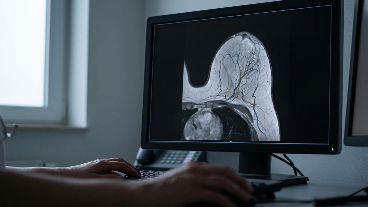
MIP reconstructions of contrast-enhanced breast MRI are a common aid for reviewing breast tissue quickly, because they condense the enhancement of every slice into a single image. Their weak point is well known: blood vessels also take up contrast and appear as bright streaks crossing the tissue, which can be mistaken for lesions or partly conceal them.
What this work adds
The authors present DeepVEST, a deep learning method trained to segment vessels in breast MR images and remove them from MIPs. Development used a retrospective analysis of scans from the public Duke-Breast-Cancer-MRI dataset. Validation went beyond automated metrics: a reader study was organised with five breast radiologists, who assessed images with and without vessel removal drawn from two datasets, Duke-Breast-Cancer-MRI and Advanced-MRI-Breast-Lesions.
Main results
Automatic vessel segmentation reached a Dice similarity coefficient of 0.611 against manual annotations, a moderate performance that the authors themselves frame as a starting point. The reader study collected 150 assessments and 600 responses. In 91 of the 150 assessments (60.7%) vessels partially or fully obscured lesion margins. Vessel removal effectiveness was rated 3.820 ± 0.749 on a 5-point Likert scale. Artifacts appeared in 45 of the 150 assessments (30.0%), but with a very low mean severity of 0.493 ± 0.900 on a 0-5 scale, indicating a minimal effect on image quality. Interreader agreement was substantial both for removal quality (Gwet coefficient 0.703) and for artifact evaluation (0.736).
What it means
This is a post-processing tool, not a diagnostic system: it does not decide whether cancer is present and does not replace the radiologist’s reading. Its value lies in removing one specific source of difficulty in challenging cases, those in which a vessel overlaps the lesion border and forces the reader to check several planes to be sure. Open questions remain: the Dice of 0.611 leaves room to improve mask accuracy, and artifacts, although mild, appear in nearly a third of images.
Reading from the breast unit
MIPs are part of routine work in many units using breast MRI, particularly for staging and for surveillance of high-risk patients. An automatic aid that cleans the image before reading could save review time and support double reading, provided it is validated on scanners and protocols other than the public datasets used here and that radiologists keep access to the original images.
Reference: Cantone M, Zhang T, Marrocco C, Han L, Portaluri A, van Dijk J, et al. DeepVEST: Deep Learning-based Vessel Segmentation and Erasure on Breast MR Images for Improved Lesion Assessment. Radiology: Artificial Intelligence, 2026. DOI